Formation of calcium oxalates and carbonates due to water stress in “nopalitos”
DOI:
https://doi.org/10.56890/jpacd.v26i.553Keywords:
Micromosaics, in situ, image overlay, water stress, Calcium Oxalates and Calcium Carbonates.Abstract
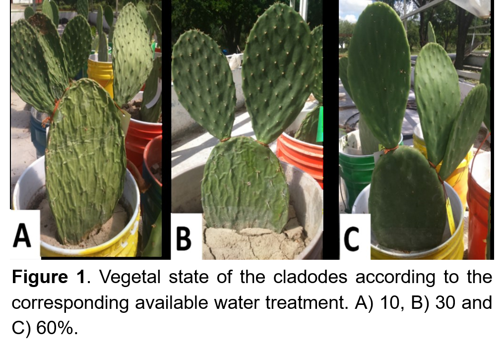
The “nopalitos” (Opuntia ficus-indica L. Miller) is an essential source of calcium in the human diet; however, the bioavailability and viability of the mineral are limited by precipitating in the form of calcium oxalate (CaC2O4) when the plant is exposed to water stress. The objective of this study was to quantify calcium oxalates and carbonates in cactus subjected to water differentials of 10, 30 and 60% of available water (AW) through photomicrographic analysis. The micro mosaics are composed of sequential images of a 10x amplitude, taken in a petrographic microscope with different light sources, plane-polarized light (LPP), cross-polarized light (LPC), and light compensated (LPC). High spectral resolution mosaics were produced using geospatial operators (ArcGIS v.10.1 and ERDAS Imagine, 2014v®). The CaC2O4 and CaCO3 were identified by spectral signature or brightness degrees in RGB format images, quantifying the minerals and surface they occupy in the cladode. The quantification of minerals in situ in nopal cladodes at different water stresses showed significant differences (p<0.05) in the area occupied by oxalates at 10% water stress and calcium carbonates at 60% within the vegetal structures of nopal vegetables. Consequently, using high-resolution mosaics and spatial operators allows the identification and quantification of mineral biomarkers in plant tissues in situ. Therefore, using image overlay, the proposed method is an alternative to the in situ quantification of minerals in plant tissues.
Publication Facts
Reviewer profiles N/A
Author statements
Indexed in
- Editor & editorial board
- profiles
- Academic society
- Journal of the Professional Association for Cactus Development
- Publisher
- Professional Association for Cactus Development

To learn about these publication facts, click ![]()
PF is maintained by the Public Knowledge Project